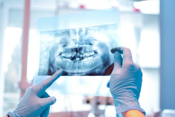
 Dental X-rays are tools used in general dentistry to help patients and providers get a better understanding of the health of the teeth and mouth. These are considered both diagnostic and preventative resources, and patients experience little discomfort for the procedure. There are a variety of dental X-ray options, as each has a different purpose and can present different information to a dental provider.
Need for X-rays
Images of the teeth provide a better picture of a patient’s oral health. Low levels of radiation from the X-rays take a picture of what is happening inside the teeth or gums. These images reveal impacted teeth, cavities, and tooth decay. Dental X-rays can show several things:
- Damages in fillings
- Signs of bone loss
- Spots of decay hidden between the teeth
- Signs of cysts or cancer
- Infections or complications with the nerve of a tooth
- Development of normal or impacted teeth
General dentistry appointments are used to assess current oral health but also to look for early signs of teeth or gum conditions. Looking inside the teeth or below the gums presents a more comprehensive view than an oral exam.
X-ray process
The patient is seated upright in a chair and covered with a lead apron or protective collar around the neck to minimize radiation exposure. Depending on the type of X-ray chosen by the dentist, the film or X-ray sensor will be fitted into a section of the mouth. It is typically the placement of the sensor that could cause some discomfort, but digital X-rays are slowly replacing these awkward film sensors.
Types of X-rays
There are different options with X-rays, as the resulting images reveal specific things about the teeth or gums. A dentist may recommend one or more of the following based on the symptoms or signs a patient exhibits.
Bitewing X-rays
These images are often recommended as a yearly procedure to check for cavities between the teeth or to review the level of bone beneath the gums. These provide a base assessment to look for changes from year to year.
Occlusal X-rays
While not used as often as a bitewing or other forms, these X-rays provide detailed images of the floor or roof of the mouth. A dentist may use this picture to look for impacted teeth, extra teeth, jaw issues, growths, or other abnormalities.
Periapical X-rays
This image presents a full picture of the tooth from the top of the crown down to the top of the root. This X-ray is often recommended when a patient has symptoms with a particular tooth or to follow up on an oral procedure. It can detect deep decay, problems within the bone structure, or an abscess.
Panoramic X-rays
This imaging is generally used every three to five years for an overall mouth evaluation. It could also be recommended prior to an oral or orthodontic procedure.
Conclusion
Dental X-rays usually have little physical impact on a patient and are not considered painful. The images are used to support general dentistry evaluations of the teeth and gums.
Request an appointment or call Midtown General & Cosmetic Dentistry at 704-307-4525 for an appointment in our Charlotte office.
Related Posts
Bad breath can be easily addressed by general dentistry practices — but some patients try to hide the problem with strongly flavored toothpaste, mouthwashes, or breath mints. Bad breath can represent a problem with oral health, and it sometimes points to significant gum disease. Learn more below about the causes of bad breath and how…
Many people have anxiety regarding going to the dentist. General dentistry visits can be easier if you have some tools to ease your nerves and get through your appointment with minimal stress. Fortunately, there are ways to ensure that you are calmer during dental or other medical appointments.A significant number of people fear going to…
Good oral health does not just require strong, clean teeth; it is the result of numerous tissues and systems working together. Saliva plays a particularly important role in general dentistry. This fluid is essential for a healthy mouth and body.The human body is continuously producing saliva. In a healthy adult, it is normal to create…
